This content includes a lot of grammatical and vocabulary errors.Please cut me some slack from Japan.
I will just describe the stimulation sites and one-point advice.
This time, we will focus on three typical nerves in the lower limb.
Tibial nerve
CMAP
・Recording electrode: abductor pollicis brevis muscle belly
・Reference electrode: on the MTP joint of the thumb
・Proximal stimulation: 7 cm proximal to the recording electrode. Proximal stimulation: 7 cm proximal to the recording electrode.
・Distal stimulation: middle of the knee socket

The tibial nerve is felt stiffly around the point where the line connecting the endocondyle and calcaneus divides 1:2. Or the midpoint between the endocondyle and Achilles tendon, but this is slightly deeper and harder to touch.
・Push the stimulating electrode in as hard as you can when stimulating at the knee socket. The back of the knee is full of meat, so if you don’t press down on it, you won’t get much out. Also, be aware that the peroneal nerve will react if it is on the outside or closer to the fibula.
Peroneal nerve
CMAP
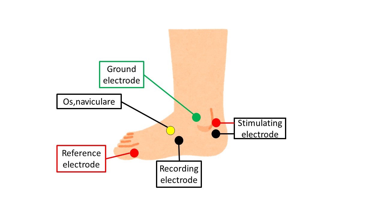
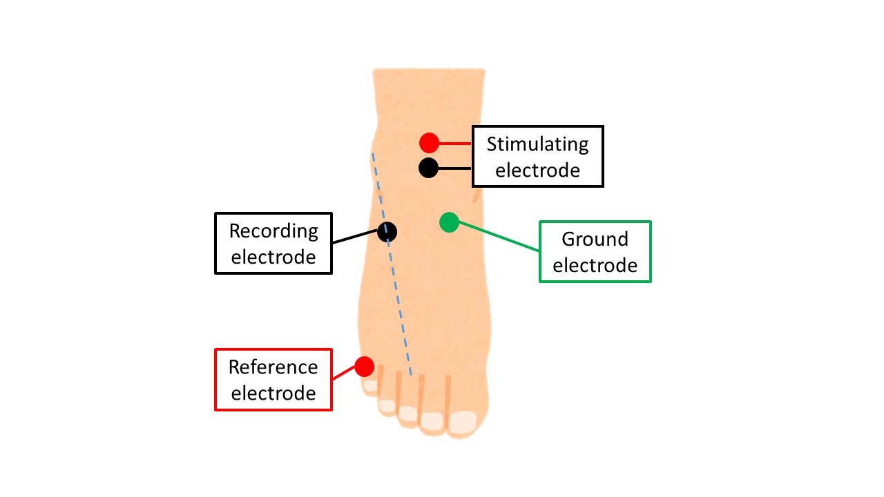
・Recording electrode: extensor digitorum brevis
・Reference electrode: on the MTP joint of the little finger
・Proximal stimulation: 7 cm proximal to the recording electrode. Proximal stimulation: 7 cm proximal to the recording electrode.
・Distal stimulation: “below” the head of the fibula and at the popliteal fossa

The EDB to which the recording electrode is attached is small, and if the disorder progresses, it will lose its shape and nothing will be touched. So, at that time, we place it at the point where the line segment connecting the middle toe and the external condyle is divided 1:2 (from the internal condyle), as shown in the figure above. It is usually there.
When stimulating with the fibular head, always stimulate with the fibular head “below” the fibular head. This allows us to examine the peroneal head, where the peroneal nerve is most likely to be injured, when we next stimulate the popliteal fossa.
When stimulating the popliteal fossa, we stimulate from the center of the fossa to slightly lateral and closer to the fibula. The long head tendon of the biceps femoris muscle is clearly visible nearby, so sometimes it is easier to stimulate in the vicinity of the tendon.
Peroneal nerve
SNAP
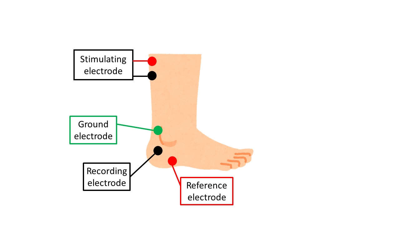
・Recording electrode: midpoint of the line connecting the calcaneus and the external condyle
・Reference electrode: 3 cm distal to the stimulating electrode
・Stimulation electrode: 14 cm proximal to the recording electrode at the lateral border of the Achilles tendon

If the waveform is difficult to obtain, it is easier to obtain it when the patient is in the supine or prone position. The peroneal nerve is a sensory nerve in the lower extremity, and is therefore very susceptible to neuropathy and edema. For this reason, the peroneal nerve is the most frequently affected by neuropathy and edema. Do not be discouraged if you cannot get it out.
That is all. Thank you again.

