This content includes a lot of grammatical and vocabulary errors.Please cut me some slack from Japan.
In this article, we will discuss three typical nerve stimulation methods and electrode positions for the upper extremity.
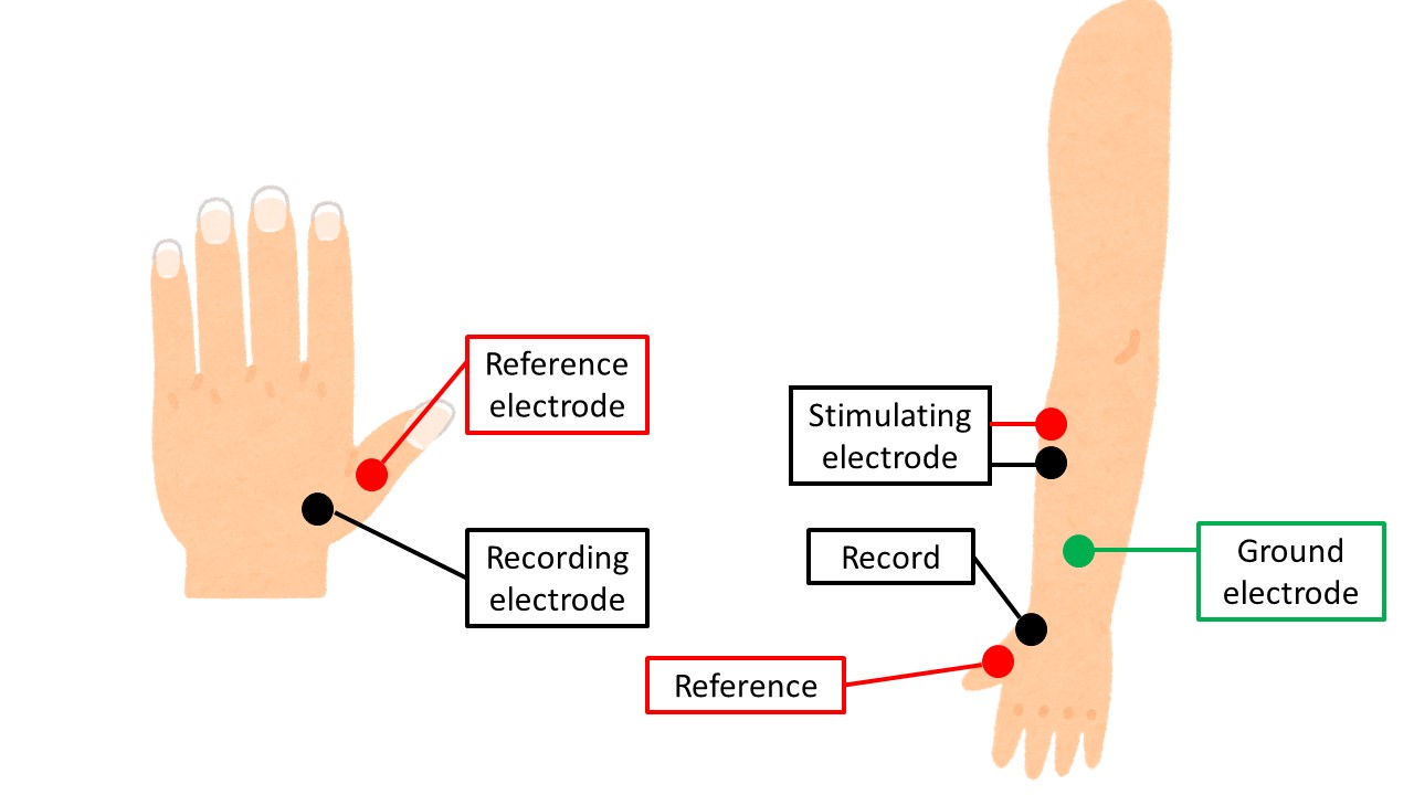
Median nerve
CMAP
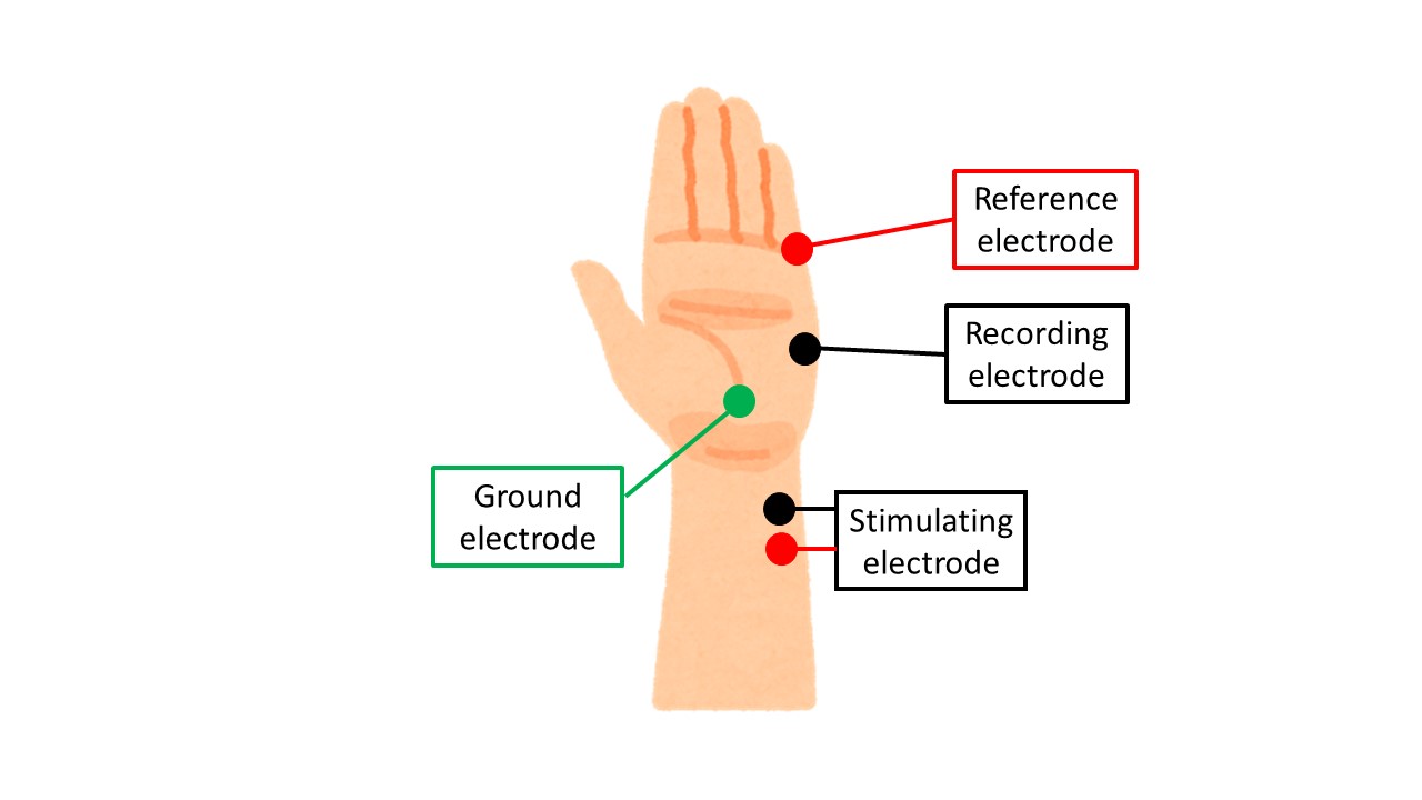
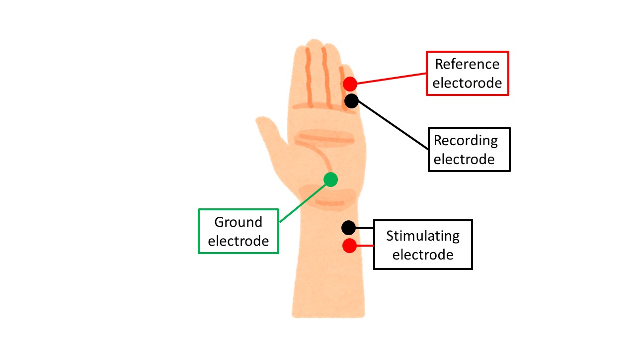
・Recording electrode: abductor pollicis brevis (APB) muscle belly
・Reference electrode: on the MP joint of the thumb
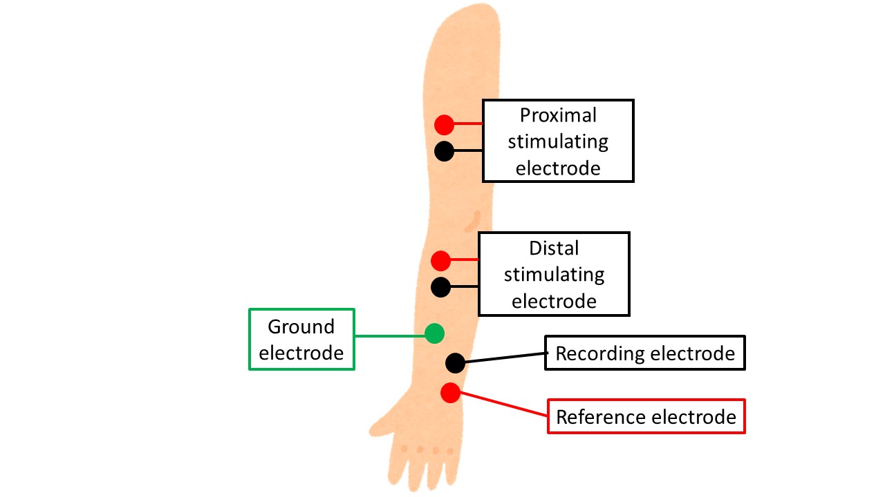
・Proximal stimulation: 7 cm proximal to the recording electrode. Proximal stimulation: 7 cm proximal to the recording electrode, between the palmaris longus tendon and the flexor digitorum tenosynovitis tendon.
・Distal stimulation: medial side of biceps tendon at elbow joint. Medial 1/4 of elbow joint, quite medial.

The long palmaris longus muscle is clearly visible when the wrist is flexed with all fingers attached. The flexor carpi radialis tendon is located on its lateral (flexor) side, and the median nerve is located between the tendons of the two muscles.
The proximal nerve is located about 1/4 of the way up the elbow joint. The brachial artery runs next to the median nerve, so it is best to place the stimulating electrode near the brachial artery. It is also easier to stimulate the biceps tendon from the inside of the elbow joint by pressing the electrode against the tendon from below.
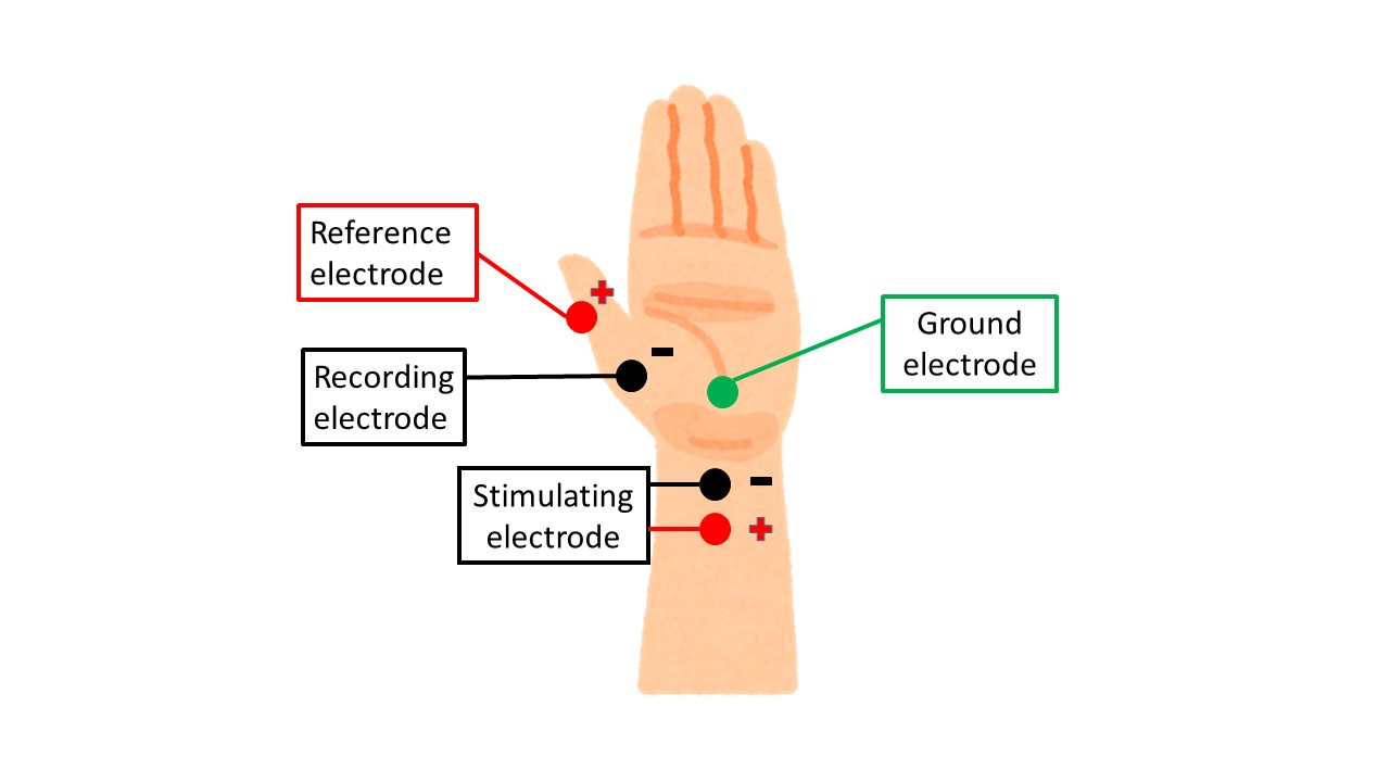
SNAP
Recording electrode: on the PIP joint of the index finger
Reference electrode: on the DIP joint of the index finger
Stimulation electrode: 14 cm proximal to the recording electrode. Between palmaris longus tendon and flexor digitorum tenosynovitis tendon

Ulnar nerve
CMAP
Recording electrode: Abductor digiti minimi (ADM) muscle belly
Reference electrode: on the MP joint of the little finger
Proximal stimulation: 7 cm proximal to the recording electrode. Proximal stimulation: 7 cm proximal to the recording electrode, just below the ulnar carpometacarpal flexor tendon
Distal stimulation: above the medial epicondyle- olecranon line, just below the medial epicondyle.
During distal stimulation, the stimulating electrode should be placed ulnarly under the ulnar carpometacarpal flexor tendon.
For proximal stimulation, inching may be used. The reference point at the elbow is the straight line between the medial epicondyle of the humerus and the olecranon. 3 cm distal to the reference point is marked as BE30 (Below Elbow 30mm, 30mm below the elbow), and 3 cm, 6 cm and 9 cm proximal to the reference point are marked as AE (Above Elbow) 30, AE60 and AE90 respectively.
The point is that the more proximal the stimulation position (AE30 => 60 => 90), the more upward from the elbow toward the biceps brachii.

SNAP
Recording electrode: on the PIP joint of the little finger
Reference electrode: on the DIP joint of the little finger
Stimulation electrode: 14 cm proximal to the recording electrode. Lateral and flexor aspect of the ulnar carpal flexor tendon
Radial nerve
CMAP
Recording electrode: Extensor indicis propria (EIP) muscle belly
Reference electrode: ulnar eminence
Proximal stimulation: 7 cm proximal to the recording electrode. Proximal stimulation: 7 cm proximal to the recording electrode, middle to slightly ulnar side of the upper arm
Distal stimulation: Upper arm between deltoid tendon and triceps muscle. Radial nerve groove
The EIP (extensor indicis brevis muscle) to which the recording electrode is attached originates from the ulna, so the electrode should be attached around the ulna as well. Note that it is often attached to the radius.
Distal stimulation is performed slightly ulnar to the middle of the upper arm, touching the ulna if the patient is not very good at it.
Proximal stimulation is difficult. I personally stimulate between the lateral humeral surface (where the deltoid attaches to the humerus) and the triceps, or at the midpoint between the acromion and lateral epicondyle with the stimulating electrode pointed along that line. It is located more proximal and lateral to the humerus than one might expect.
SNAP
Recording electrode: anatomical tobacco socket
Reference electrode: MP joint of the thumb
Stimulation: 14 cm proximal to the recording electrode. Stimulus: 14 cm proximal to the recording electrode.
The recording electrode is placed in the anatomical tobacco field, and the reference electrode is placed 3 cm distal to the MP joint of the thumb.
Stimulation is more likely to occur if the electrode is applied to the radius from above or below the radius (on the palmar side).
Please refer to the next article for the typical nerves of the lower limb.
That is all. Thank you again.