This content includes a lot of grammatical and vocabulary errors.Please cut me some slack from Japan.
In this article, we will explain the waveforms of needle electromyography and their significance, focusing on the most important ones: Positive sharp wave (P wave), Fibrillation (spontaneous potential of fibers), Fasciulation (spontaneous potential of fiber bundles), and Polyphasic wave (polyphasic wave).
Positive sharp wave
& Fibrillation (commonly known as p/fib)
These are all important findings that can be seen at rest and suggest that “the disorder is occurring right now and may be cured if treated immediately. It is no exaggeration to say that these are the most important findings of the needle EMG, or rather, this is what we are doing.
Both P wave and Fib reflect “fascial instability” and are seen in both muscle diseases (myositis, etc.) and nerve diseases (demyelination, conduction block).
Positive sharp wave is a monophasic wave that plunges down and rises up slowly, as shown in the figure below. Fibrillation, on the other hand, is a wave of relatively short duration that rises rapidly after a sudden drop.

In fact, the Positive sharp wave and Fibrillation are the same wave, but they appear to be different only because of the location of their occurrence and the position of the recording site. Therefore, they are synonymous and can be observed at the same time.
If you watch or rather listen to the video below, you will hear a “popping” sound as if it is raining. Please remember that this is the sound of Positive sharp wave and Fibrillation. But it is actually a higher pitched sound, like “Totata-tata-tata! sound like rain hitting a tin roof. This video is a little far away from the sound, so you should get a little closer. Bitch.
Video Link: P wave + Fibrillation
Occasionally Positive sharp wave/Fibrillation and normal voluntary contraction waveforms look similar. The way to distinguish between them is that Positive sharp wave/Fibrillation is similar to involuntary movement, which is instability of fascia, so it tends to appear periodically. On the other hand, normal waveforms appear randomly because the patient’s consciousness (voluntary contraction) is involved.
Fasciculation
Another waveform seen at rest is Fasciculation (fibrofibrillatory spasm). Fasciculation is a condition in which the brain does not give a command to the muscles to contract, but the nerves run wild on their own (spontaneous discharge of anterior horn cells and axons, to be precise).
Video link: Fasciulation from about 11 seconds in.

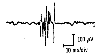
It looks very similar to the Polyphasic wave (described below), with the only difference being a smaller potential of 0.2-0.8mv.
The sound is characteristically lower than that of Positive sharp wave and Fibrillation. Fasciulation can also be seen in normal conditions, so its presence does not necessarily mean that it is abnormal, but it can help in the diagnosis of suspected neurological diseases such as ALS.
Fasciulation can be seen in both acute and chronic phases, so it is not used to determine the extent of the disease.
Polyphasic wave
In Japanese, it is called “polyphasic wave.
Unlike the other three, this is a finding that is seen during voluntary contractions. When we find it, we judge that “time has passed since the disorder occurred, and that it is in the chronic phase. Especially when there is a huge wave with a potential of over 5 mv, I judge that it has been a long time and that recovery will be difficult.
Polyphasic waves are similar in shape to fasciulation but different in size. Polyphasic waves usually have a potential of 2 mv or more and a duration of 10 ms or more.

Video link: Polyphasic wave
This polyphasic wave can be seen in normal people over the age of 60. This is because some nerves die with aging. Therefore, polyphasic waves are considered significant only when differences are observed in the same muscle and multiple locations in elderly patients.
That’s all. Thank you again.

